Work in Progress: Developing a Screening Test for Condylar Fracture Risk in Horses
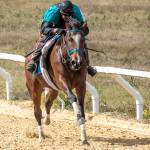
Although rare, stress fractures of the cannon bone can lead to catastrophic injuries, especially in Thoroughbred racehorses. Having a screening test to assess the risk of stress fracture would help prevent injuries, thus improving welfare. A preliminary study using both digital radiographs and standing computed tomography found tomography to be the better tool for detecting bone changes that may predispose horses to serious or catastrophic injuries.*
Stress fractures leading to full condylar fractures and possible catastrophic injury occur due to progressive accumulation of fatigue microdamage of the subchondral bone at the end of the cannon bone. Being able to detect the microdamage during a routine screening test will help prevent injury. However, the anatomy of that region of the cannon bone is complex, making screening challenging.
In a study by Irandoust and coworkers, 31 limbs from Thoroughbred racehorses were imaged using both digital radiography and standing computed tomography. Four experienced observers evaluated each of the images for the presence and extent of dense bone as well as lucencies and fissures. They also assigned a risk assessment grade for condylar stress fracture. The sensitivity (few false negatives), specificity (few false positives), and risk of fracture were compared to a reference standard.
“Calculating the sensitivity and specificity is important because a good screening test must be accurate. Too many false negatives result in injuries, and too many false positives will result in horses being needlessly scratched from races,” advised Kathleen Crandell, Ph.D., a nutritionist for Kentucky Equine Research.
Standing computed tomography was better able to detect subchondral structural changes of the cannon bone and risk assessment for condylar stress fractures than digital radiography. Specifically, sensitivity for detecting structural changes was lower than specificity for both digital radiography and standing computed tomography, and sensitivity for risk assessment was lower than specificity for all four observers. Sensitivity was improved with standing computed tomography, especially in horses classified as having a high risk of injury compared to the reference assessment.
Additional studies are needed to apply these findings to racing Thoroughbreds.
“Even for condylar fractures of the cannon bone that are successfully repaired surgically, future athletic performance can be disappointing because of the development of osteoarthritis,” Crandell said.
Osteoarthritis can develop following any type of trauma involving a joint due to inflammation affecting all joint tissues and the subsequent destruction of articular cartilage.
“For the daily wear and tear that can create inflammation in the joints of an athletic horse, joint supplements can be used prophylactically, prior to overt trauma, to prevent OA. Well-formulated supplements aid in maintaining joint integrity, reducing cartilage damage, and stimulating cartilage repair,” said Crandell.
*Irandoust, S., L.M. O’Neil, C.M. Stevenson, F.M. Franseen, P.H.L. Ramzan, S.E. Powell, S.H. Brouts, S.J. Loeber, D.L. Ergun, R.C. Whitton, C.R. Henak, and P. Muir. 2024. Comparison of radiography and computed tomography for identification of third metacarpal structural change and associated assessment of condylar stress fracture risk in Thoroughbred racehorses. Equine Veterinary Journal:14131.

