Ultrasound Reliably Assesses Cartilage Thickness in Horses
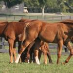
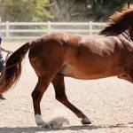
The fetlock joint, also called the metacarpophalangeal joint, lies between the cannon bone and the first or long pastern bone in the lower forelimb. The fetlock joint is a common location of osteochondrosis, a developmental orthopedic disease characterized by cartilage flaps, fragments, and cysts in the underlying bone. Assessing cartilage in young foals is therefore crucial.
Causes of osteochondrosis are unclear but believed to be multifactorial, including genetics, rapid growth rate, anatomic characteristics, nutrition, and trauma. In general, osteochondrosis is attributed to a “failure of endochondral ossification” secondary necrosis within the cartilage during growth.
The most common site of osteochondrosis in foals is the distal third metacarpal bone, the cannon bone, within the fetlock joint. More specifically, osteochondrosis is frequently localized to a specific region called the sagittal ridge.
Many osteochondrosis lesions can be diagnosed on X-ray, which is why X-rays are often part of presale or prepurchase exams. These X-rays, however, rely on secondary signs of articular cartilage damage, including subchondral bone changes (the layer of bone lying directly under the cartilage) and osteoarthritis changes.
In contrast, ultrasonography can directly visualize the cartilage, and studies show that ultrasound is superior to radiography for identifying cartilage lesions in various joints in adult horses.
“If ultrasound can assess articular cartilage thickness in juvenile horses, then this noninvasive imaging modality would be a valuable tool in equine practice,” said Kathleen Crandell, Ph.D., a Kentucky Equine Research nutritionist.
To evaluate if ultrasound can reliably assess cartilage thickness of the distal cannon bone in the fetlock joint, 33 fetlocks from foals 12 days to 10 months old were evaluated.* Cartilage thickness measurements obtained via ultrasonography were compared to the cartilage thickness measured microscopically and using computed tomographic arthrography (CTA, described previously as the most accurate way of identifying cartilage lesions in adult horses).
In this population of foals, cartilage thickness measured by CTA agreed poorly with direct measurement of cartilage thickness using microscopy. There was better, though still weak, agreement between cartilage thickness measured ultrasonographically compared to direct microscopic measurement. However, the best agreement was at the sagittal ridge of the canon bone—probably the most important site of the fetlock where osteochondrosis commonly occurs.
“This study shows that ultrasound is more accurate than CTA in assessing cartilage thickness in the third metacarpal bone of young horses,” explained Crandell. “Further, the sagittal ridge of the third metacarpal bone was reliably assessed by ultrasound.”
The researchers therefore concluded that ultrasound screening of the juvenile cannon bone for assessing cartilage and the cartilage-bone interface is a reliable technique. The best agreement of cartilage thickness occurred at the sagittal ridge where osteochondrosis lesions most commonly occur.
According to Crandell, “There is only so much that can be controlled in preventing the development of osteochondrosis in foals because it is a multifactorial disease. From a nutritional perspective, keeping the diet moderate in nonstructural carbohydrates and moderate in quantity as well as supplying optimal amounts of nutrients in balance with each other are key.”
DuraPlex is a proprietary blend of specific proteins, minerals, and vitamins scientifically proven to increase bone mineral density and bone area in growing horses. It supports bone growth by providing the nutrients necessary for strong bone development and preventing demineralization of bone during stall rest or when limited turnout is available.
*Hoey, S., U. Fogarty, H. McAllister, A. Puggioni, B. Cloak, H. Richard, C. Skelly, and S. Laverty. 2025. Ultrasonographic assessment of equine metacarpal cartilage thickness is more accurate than computed tomographic arthrography. Veterinary Radiology and Ultrasound 66(1):13444.
Most Popular
 Putting Weight on a Skinny Horse (350,339)
 Benefits of Beet Pulp for Horses (238,593)
 Hot Blood, Warm Blood, Cold Blood in Horses (183,785)
 Possible Link Between Selenium and Cribbing in Horses (171,952)