Feeding, Exercise, and Blood Flow Distribution in Horses
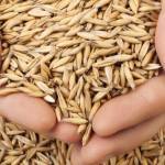
All horses must eat, and most horses must perform some type of work. A complex relationship exists between the type and amount of feed given, the duration and intensity of work expected of the horse, the impact of feeding on the digestive system, and the impact of exercise on gut function and nutrient digestibility.
Anyone who has fed horses realizes that these animals have a profound cardiovascular response to feeding. The noise associated with rattling buckets in a feed room can send a stable of horses into a frenzy. This phase of feeding is known in the scientific literature as the anticipation/ingestion phase. The anticipation/ingestion phase begins when the animal becomes aware of an upcoming feeding and continues during ingestion of food, gradually decreasing after the food has been eaten. In ponies, the cardiovascular response to feeding remains elevated for more than one hour following consumption of a grain meal.
Along with cardiovascular changes in response to feeding, changes can also occur in the distribution of blood flow within the body, which is controlled centrally through the nervous system and locally by the environmental conditions in the immediate vicinity of the blood vessels. Central control of blood flow is mediated by the autonomic nervous system, primarily by the sympathetic nerve fibers. Increased sympathetic nerve activity (excitement/panic) causes constriction in the arteries, sending blood to the abdominal organs and causing dilation of arteries in skeletal muscle. This scheme shunts blood away from the digestive system to working muscles in an adrenaline-driven flight response.
Local control of peripheral blood flow appears to be adjusted to the existing metabolic activity of the tissue. According to the metabolic model of tissue blood flow, any intervention that results in an oxygen supply that is inadequate for the requirements of the tissues gives rise to the formation of vasodilator metabolites. These metabolites are released from the tissue and act locally to dilate the smaller arteries, increasing the flow of blood and the supply of oxygen.
Blood is distributed and redistributed to the various tissues of the body by arteriolar smooth muscle control. In fasted ponies (24-hour fast), 20.4% of cardiac output (blood flow) is distributed to the various tissues of the digestive system, while 79.6% of the blood flow is found in other tissues, including skeletal muscle. Once a horse is fed, the amount of blood distributed to the gastrointestinal tract increases. This mesenteric hyperemia is confined to the digestive organs actively engaged in digestive functions. The net result of feeding in horses is a redistribution of cardiac output such that 27.4% of blood flow is distributed to the various tissues of the digestive system, while 72.6% of the blood flow is found in nondigestive tissue.
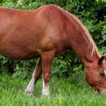
Limited information exists regarding changes in blood flow distribution during exercise in fed compared with fasted horses. Blood flow distribution during exercise was measured in
eight fed ponies and eight fasted ponies, with the fasted group not receiving feed during the 24 hours preceding data collection. During this 24-hour period, these ponies were housed in a stall containing water and salt, but devoid of bedding material. The fed treatment group was provided free-choice alfalfa hay, water, and salt during the 24 hours preceding data collection. In addition, these ponies consumed a pelleted grain concentrate at a rate of 0.7% of body weight 1.4 hours preceding data collection. Each group of ponies was monitored at rest and during 30 minutes of treadmill exercise. The treadmill was positioned at a 7% incline and set at a speed of 28 km/hour such that the ponies reached a heart rate greater than 150 beats per minute. This intensity of exercise is approximately 75% of heart rate maximum.
Blood flow to the digestive tract decreased during exercise in both fasted and fed ponies; however, blood flow was consistently higher throughout exercise in fed ponies. It is speculated that fed ponies had an elevated blood flow at rest, associated with increased digestive tissue oxygen demand, and the increased sympathetic tone produced during exercise was not large enough to totally reverse this hyperemia.
As expected, blood flow to the locomotor and respiratory muscles increased during exercise in both fed and fasted ponies. However, blood flow to both locomotor and respiratory muscles was higher in fed than in fasted ponies. It is likely that fed ponies worked harder during exercise, as evidenced by increased muscle blood flow, due to increased gut fill associated with feeding. Because the fed ponies exercised at approximately 75% of heart rate maximum, they were able to increase heart rate, cardiac output and stroke volume to deliver an increased amount of blood to both the digestive tract and working muscle.
Since blood flow is shunted away from the gastrointestinal tract during exercise, what is the effect of exercise on nutrient digestibility? Studies at Kentucky Equine Research (KER) showed that exercise resulted in a small but statistically significant decrease in dry matter digestibility. In that study, exercise consisted of walking, trotting, and cantering approximately five miles per day on an inclined treadmill. It is not known if the decrease in dry matter digestibility is actually biologically significant to the animal.
Most Popular
 Putting Weight on a Skinny Horse (348,916)
- Benefits of Beet Pulp for Horses (237,456)
 Hot Blood, Warm Blood, Cold Blood in Horses (183,331)
 Possible Link Between Selenium and Cribbing in Horses (171,463)